Internal degloving injury – the Morelle-Lavalee Lesion
Sometimes the level of adherence between the playing surface and the skin is so great, that there is little shearing force at the level of the skin, more so beneath the level of the hypodermis and fascia over muscle or bone, where the layers separate and bleeding occurs into the space formed.
This is an internal degloving injury known as a Morelle Lavalee Lesion which causes bruising, a fluctuant fluid filled swelling and restriction of joint movement due to the mass effect. The fluid-filled space created is not synovial lined, in contrast to say a bursa, so the bleeding can leak out into fascial places and delayed bruising developing some distance distal to the fluctuant swelling are tell-tale differentiators from a traumatic haemorrhagic bursitis.
If left, the Morelle-Lavallee lesion may encapsulate with fibrous tissue leaving a scar-based mass which can cause restriction and symptoms and may require surgical excision. Hence, these lesions should be managed aggressively and be prepared for repeated aspiration under sterile conditions to prevent encapsulation. They will, however, recur frequently and can be very difficult to manage.
Ultrasound is an excellent modality for diagnosing the lesion acutely and the tissue plane in which it sits, but if over a joint, Ultrasound can be more difficult to differentiate from a bursa with time if the area becomes encapsulated. Diagnosis is often made in Magnetic Resonance Imaging (MRI) due to the concern for associated injury.
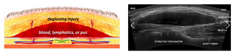
Diagram of a Morelle Lavalee Lesion in the hypodermis and ultrasound scan of similar on the hip.